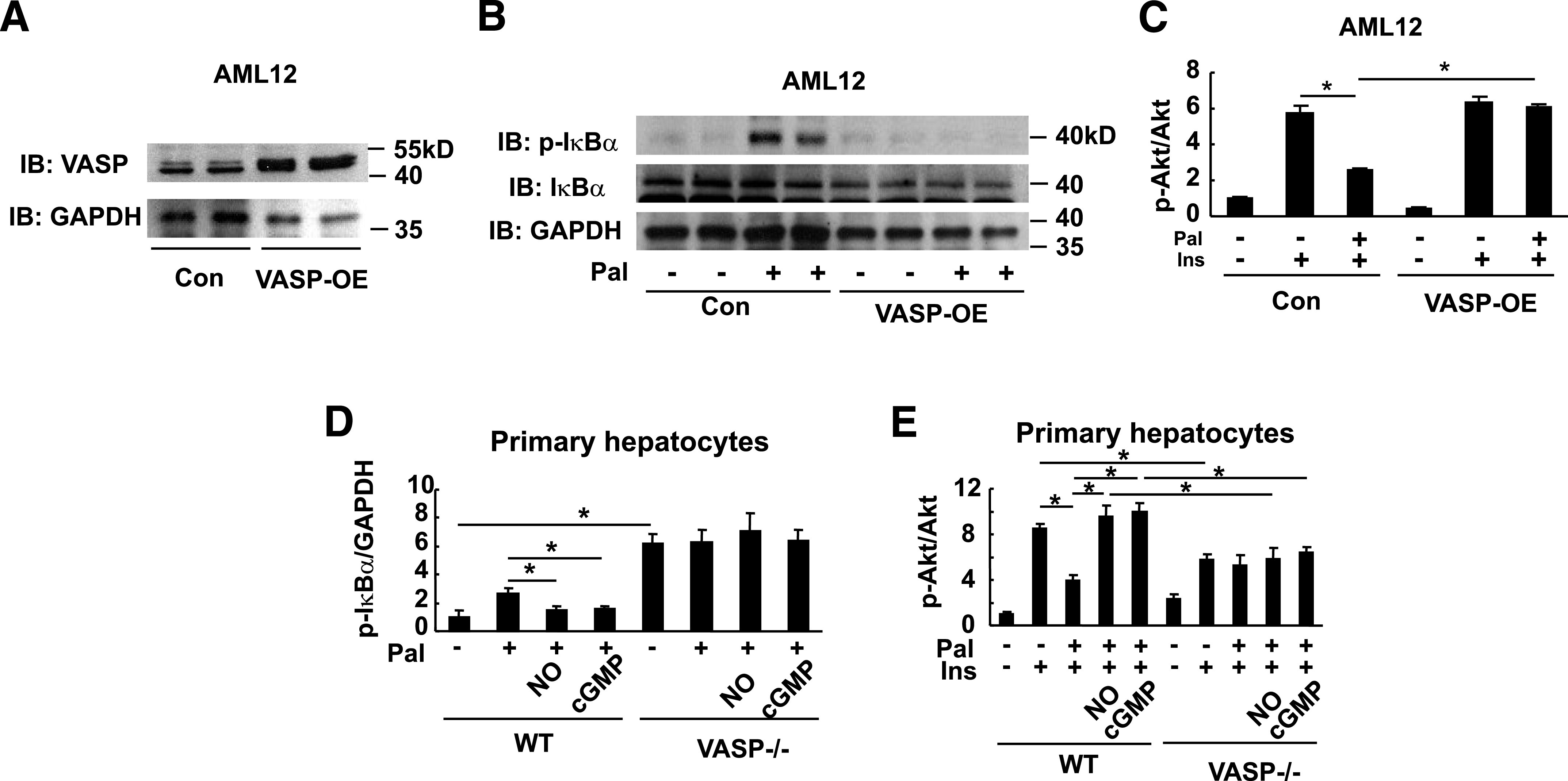
FIG. 6.

The effect of VASP signaling on hepatocyte inflammatory responses to palmitate. AML12 hepatocytes were transduced with VASP (VASP-OE) or control (Con) vector. A: VASP Western blot. B: Hepatocytes were treated with 100 μmol/L palmitate (Pal) or vehicle for 4 h, and a representative IκB-α phosphorylation Western blot from one of three independent experiments is shown. C: Insulin (Ins)-mediated pAkt signaling (10 nmol/L insulin for 15 min) in transduced hepatocytes, following 4 h palmitate (100 μmol/L). Ser 473 Akt phosphorylation was assessed by Western blot analysis, and fold increase over control condition was calculated (n = 3). *P < 0.05. D: Primary hepatocytes were isolated and cultured from Vasp−/− mice or wild-type (WT) control mice. Isolated hepatocytes were treated with palmitate (100 μmol/L) for 4 h following 4-h pretreatment with 10 μmol/L DETA-NO or 10 μmol/L 8Br-cGMP. IκB-α phosphorylation was assessed by Western blot analysis. *P < 0.05. E: Insulin-mediated pAkt (10 nmol/L for 15 min) following treatment with palmitate (100 μmol/L for 4 h), DETA-NO (10 μmol/L for 4 h), or 8BrcGMP (10 μmol/L for 4 h). Akt Ser 473 phosphorylation assessed by Western blot (n = 3). *P < 0.05. IB, immunoblot; kD, kilodalton.
