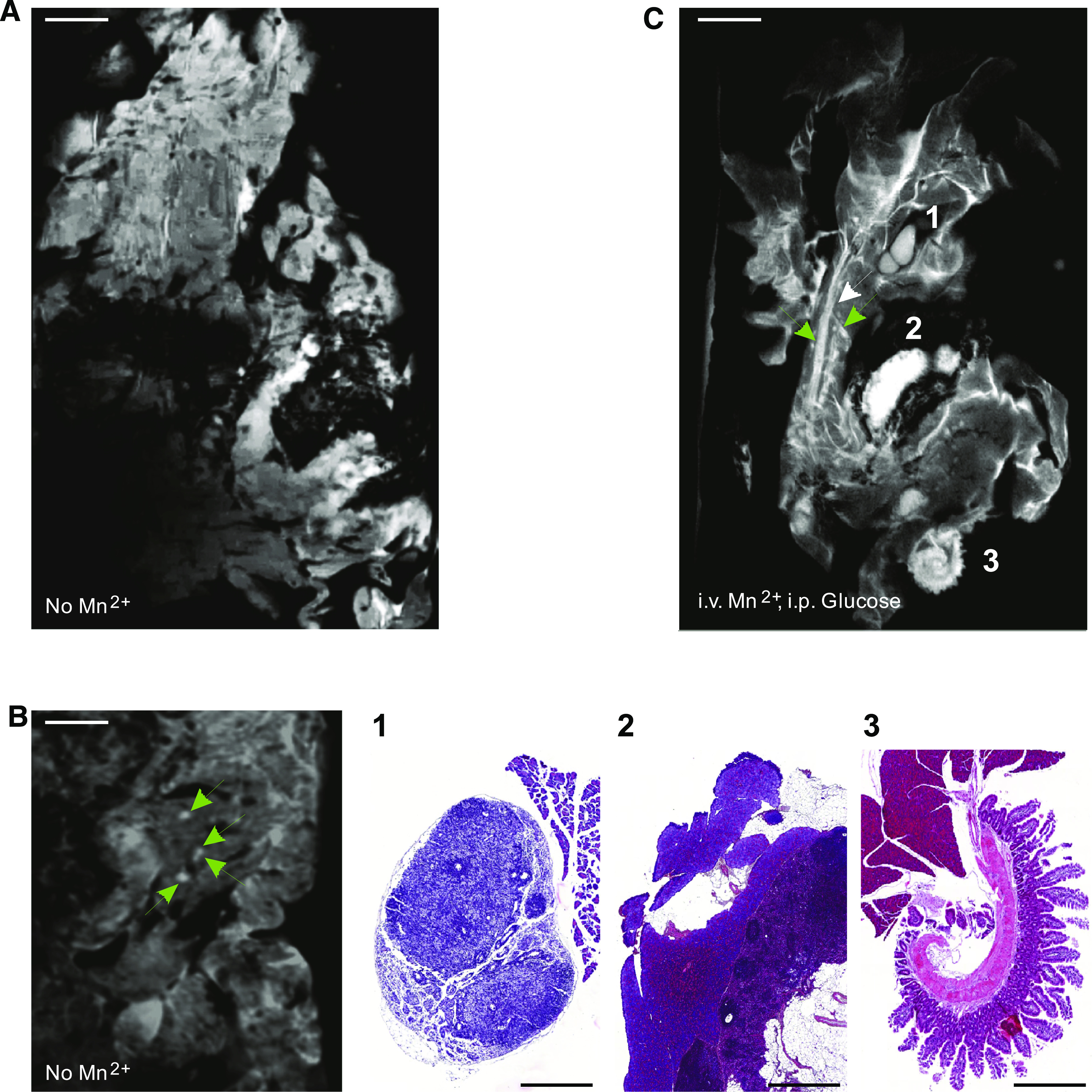
FIG. 1.

High-resolution MRI reveals the structure of the entire mouse pancreas. A and B: Whole mouse pancreata (300-μm-thick slices) were imaged ex vivo, with 50 μm in plane resolution (TR/TE = 282/7 ms). A: In an image obtained without contrast agent, pancreatic lobules are seen, most of which feature a somewhat homogeneous and structureless content at this low magnification. Scale bar: 1 mm. B: Under these conditions, MRI infrequently identified putative islets of Langerhans (green arrows) within the intact pancreas. Scale bar: 1 mm. C: The contrast of pancreatic lobules was enhanced after intravenous infusion of MnCl2 combined with an intraperitoneal injection of glucose. Under these conditions, MRI allowed to distinguish whitish tubular structures, as well as highly contrasted round-ovoid structures of various sizes. The smallest of these structures (<0.5 mm; green arrows) was observed within the pancreatic lobules. Larger structures (>1 mm) were seen between the lobules. The latter structures were identified by histological analysis of the very same pancreas, confirming that MRI differentiates the pancreatic parenchyma from intrapancreatic lymphatic ganglia (C1), spleen (C2), and loops of small intestine (C3). Scale bar: 1 mm in A–C; 0.5 mm in lower panels B1–3. (A high-quality digital representation of this figure is available in the online issue.)
