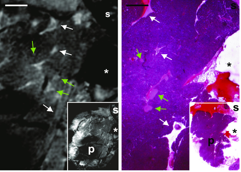
FIG. 5.
MRI detects islets in situ in a living mouse. High magnifications of MR images of the exteriorized pancreas of a living, anesthetized mouse reveal a pancreatic substructure like that observed ex vivo, including the presence of elongated vessels and ducts (white arrows) and round-ovoid bodies of small size (green arrows). Histological correlation showed that these small bodies corresponded to individual pancreatic islets. Scale bar: 1 mm. Insets show low magnification views of the same pancreas. P = pancreas; s = spleen. *Position of the plastic pin that secured the pancreas. (A high-quality digital representation of this figure is available in the online issue.)

