
FIG. 6.
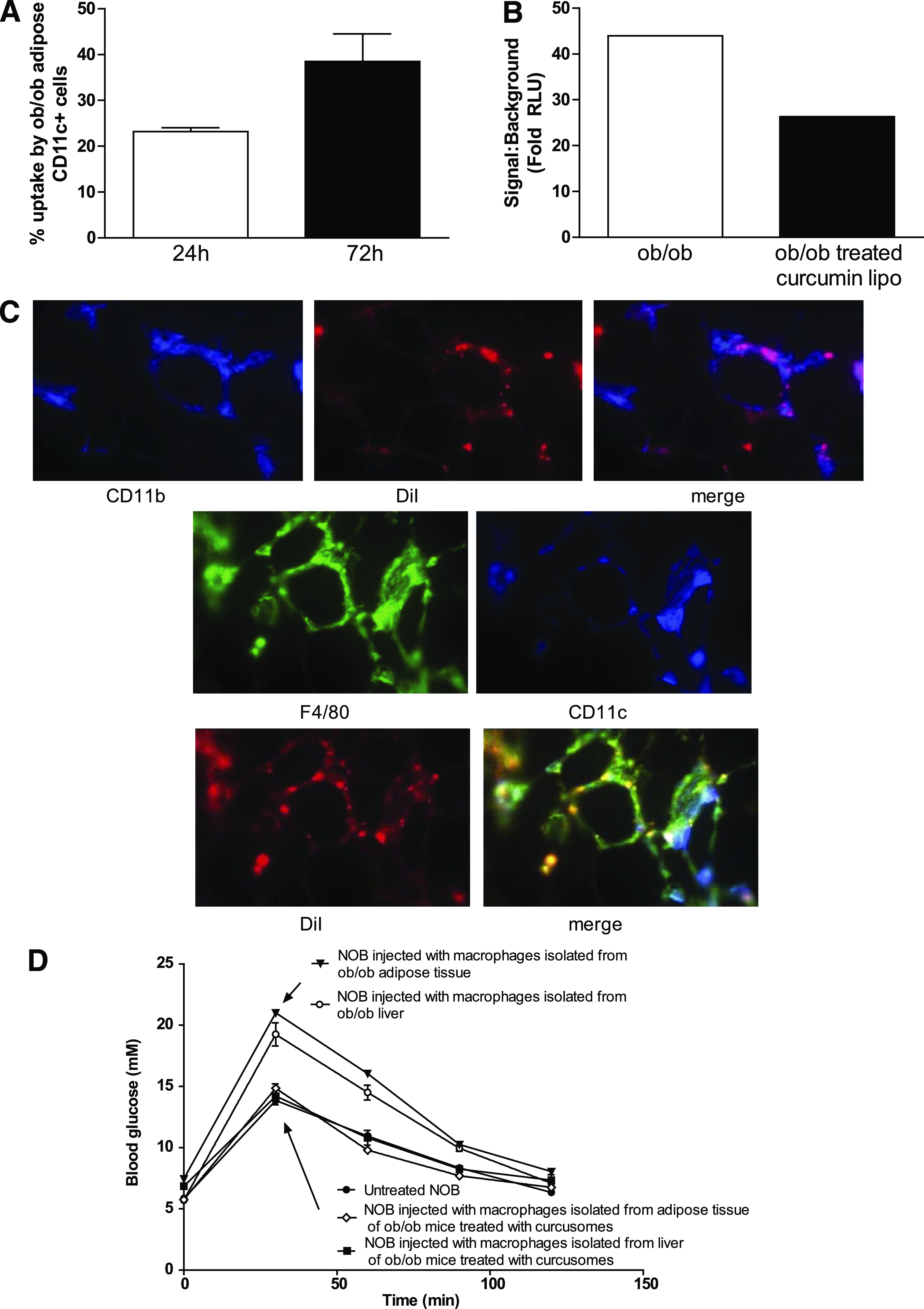
In vivo delivery of curcusomes inhibits adipose tissue inflammatory DC nuclear RelA and promotes insulin sensitivity. A: Ob/ob mice were injected intraperitoneally with DiI-curcusomes, and gradient-purified cells were harvested at 24 and 72 h and stained for MHC class II and CD11c. Shown are the percentages of CD11c+ cells that are DiI+. Data are mean ± SEM of two separate experiments. B: Nuclear extracts from CD11c+ ATM isolated from ob/ob mice injected with or without curcusomes for 24 h were analyzed for DNA binding of RelA by chemiluminescence. Shown is the mean of duplicates from two separate experiments. C: Ob/ob mice were injected intraperitoneally with DiI-curcusomes for 72 h and then adipose tissue was harvested, frozen in optimal cutting temperature media, sectioned, and stained with CD11b (blue) or F4/80 (green) and CD11c (blue) antibodies. DiI staining is indicated in red; images analyzed by immunofluorescence microscopy. Original magnification ×25. D: GTT from NOB mice injected intravenously with 1 × 10⁶ F4/80+ DCs from adipose tissue or liver tissue from ob/ob mice treated with or empty liposomes or curcusomes for 24 h. (A high-quality digital representation of this figure is available in the online issue.)