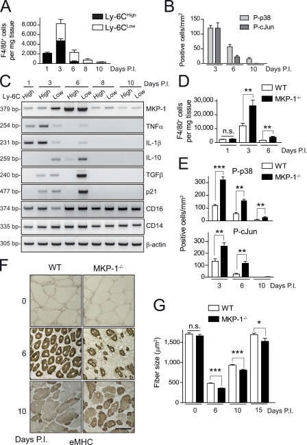
Figure 1.
p38–MKP-1 balance is required for efficient skeletal muscle repair. (A) Number of F4/80-positive macrophages in WT muscle at the indicated times postinjury (P.I.) that were obtained by flow cytometry. Total number of cells per milligram of CTX-injured muscle tissue was calculated. (B) Phospho-p38– and phospho–c-Jun (as an indicator of activated JNK)–positive macrophages (P-p38 and P-cJun, respectively) were counted in immunostained cryosections of gastrocnemius muscles obtained from WT mice 3, 6, and 10 d after injury (see pictures in Fig. S1 C). (C) Gene expression analysis by RT-PCR in isolated macrophage populations at the indicated times after injury. (D) As in A, number of F4/80-positive macrophages in WT and MKP-1−/− mice per milligram of injured muscle tissue. (E) As in B, phospho-p38– and phospho–c-Jun–positive macrophages from WT and MKP-1−/− mice at the indicated times after injury (see pictures in Fig. S1 C). (F) Muscle cryosections were stained with an anti-eMHC antibody. Bar, 50 µm. (G) As in F, cryosections were stained with H/E, and the mean area of regenerating myofibers was calculated (see pictures in Fig. S1). Means ± SEM of at least three experiments. ***, P < 0.001; **, P < 0.01; *, P < 0.05.

