
Figure c:
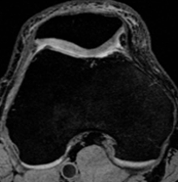
Representative MR images obtained at (a, c) baseline and (b, d) 2-year follow-up demonstrate interval progression of OA-related focal morphologic abnormalities. (a, b) Sagittal T2-weighted images obtained in a subject with a 5.1% increase in cartilage T2 over 2 years (all compartments combined, change in T2 = 1.96 msec) and a 24.5-point increase in summation WORMS. There was interval development of tearing of the posterior horn of the lateral meniscus (curved arrow in b), subchondral cyst in lateral tibial plateau (arrowhead in b), and joint effusion (straight arrow in b). The focal cartilage defect overlying the lateral tibial subchondral cyst also appeared worse in b than in a. (c, d) Transverse intermediate-weighted images from another subject with a 2.6% increase in cartilage T2 (change in T2 = 1.15 msec) and an eight-point increase in summation WORMS. At baseline (c), the patellar cartilage was essentially normal. However, there was interval development of signal heterogeneity in the lateral facet of the patellar cartilage at follow-up (arrow in d), which is suggestive of early degeneration. A trace joint effusion is also evident in d.