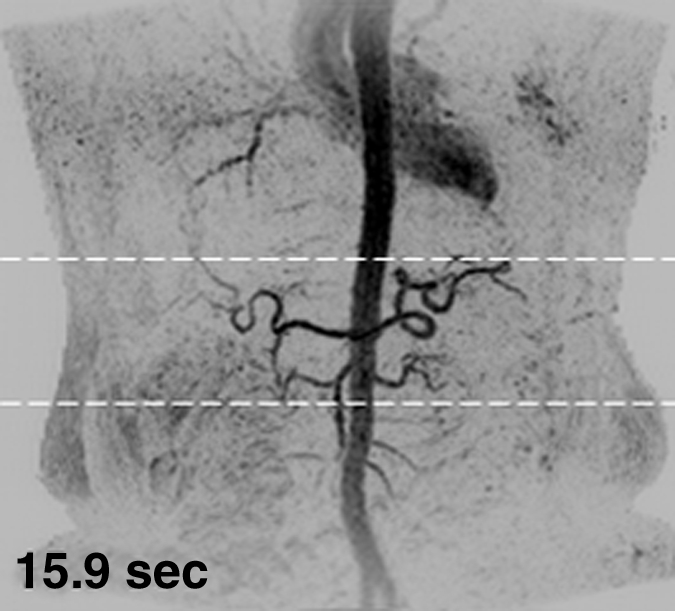
Figure 1c:

Results from the 3D timing image (3.24/1.30) with 2 mL contrast material in a 53-year-old woman (volunteer 6). (a–c) Consecutive 1.77-second coronal MIPs show progressive filling of the arterial vasculature. Time after contrast material injection is shown on each frame. (d) Targeted axial MIP from the selected superoinferior region (area between the white dashed lines in c). Axial MIP highlights the high image quality and isotropic nature of the timing image, as well as the large 3D volume that was acquired, as shown, encompassing the full superoinferior (a–c), as well as the anteroposterior and left-right (d) extent of the abdomen. Details are also in Movie 1.
