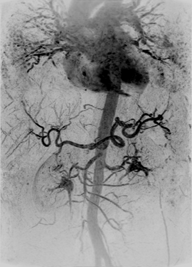
Figure 2a:

Abdominal MR angiogram and MR venogram (4.33/1.91) in a 53-year-old woman (volunteer 6). (a) Oblique MIP, 20° from coronal, of the abdominal MR angiogram shows the abdominal vasculature from the pulmonary vessels inferiorly to the iliac arteries. There is some signal intensity loss at the inferior edge of the FOV caused by coil signal drop-off. (b) Coronal MIP from the subsequent venogram shows hepatic portal and venous systems, as well as parenchymal enhancement.
