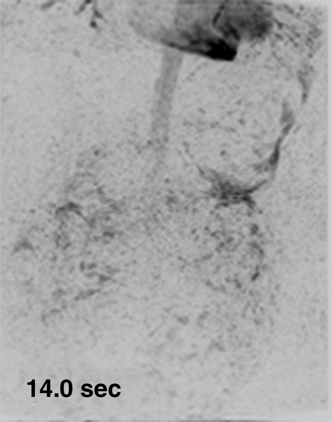
Figure 3a:

Results from examination in a 27-year-old woman (volunteer 7). (a–d) Consecutive 1.17-second coronal MIPs from the 2-mL timing image (3.09/1.23) show clear progressive arterial enhancement with high SNR, as routinely obtained. (e) Oblique MIP, 20° rotated from the coronal plane, of the renal angiogram (4.38/1.99) shows sharp detail and high SNR in the main through segmental renal arteries, with enhancement of the vessels of interest as follows: abdominal aorta, iliac arteries, renal arteries, mesenteric arteries, as well as the fine branching hepatic and mesenteric arteries. An accessory left renal artery was identified in both the timing image and angiogram (arrow), as was a replaced right hepatic artery (arrowhead) arising from the superior mesenteric artery. Details are also in Movie 2.
