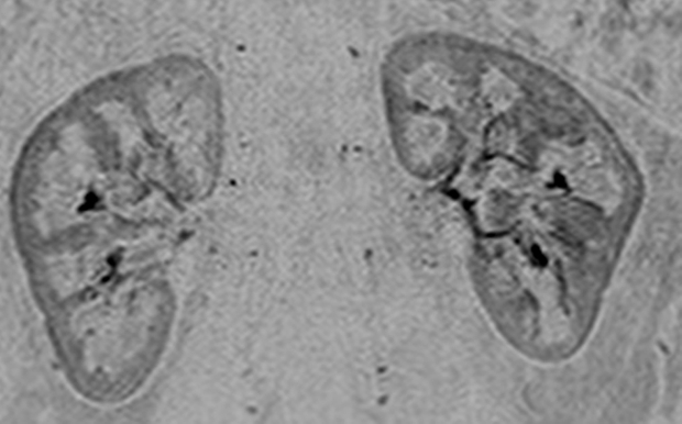
Figure 4d:

Renal angiogram (4.94/2.29) in a 36-year-old woman (volunteer 8). (a) Coronal MIP covering the full superoinferior FOV shows high SNR and fine detail throughout the abdominal arterial vasculature. (b) Targeted sagittal MIP demonstrates the visualization of the mesenteric artery origins. (c) Targeted coronal MIP about the renal arteries shows sharp delineation of the main and segmental renal arteries. (d) Single 1.60-mm-thick coronal section cropped about the kidneys shows the segmental and intrarenal arteries, as well as the enhancement of the renal cortex, that can be visualized in the source images. Details are also in Movie 4.
