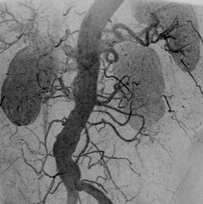
Figure 5a:

Renal angiogram (4.45/2.01) in a 63-year-old woman (patient 2). Motion artifact obscured visualization of the vasculature in every other time frame of the 3D test-bolus acquisition. Regardless, accurate timing information was obtained, which showed enhancement of the aorta and renal arteries at 31.9 seconds, more than 2 standard deviations later than the mean contrast agent arrival time of the series (18.5 seconds). (a, b) Targeted MIPs about the renal arteries and anterior mesenteric arteries illustrate the vascular detail obtained over each of these regions.
