
Figure 5f:
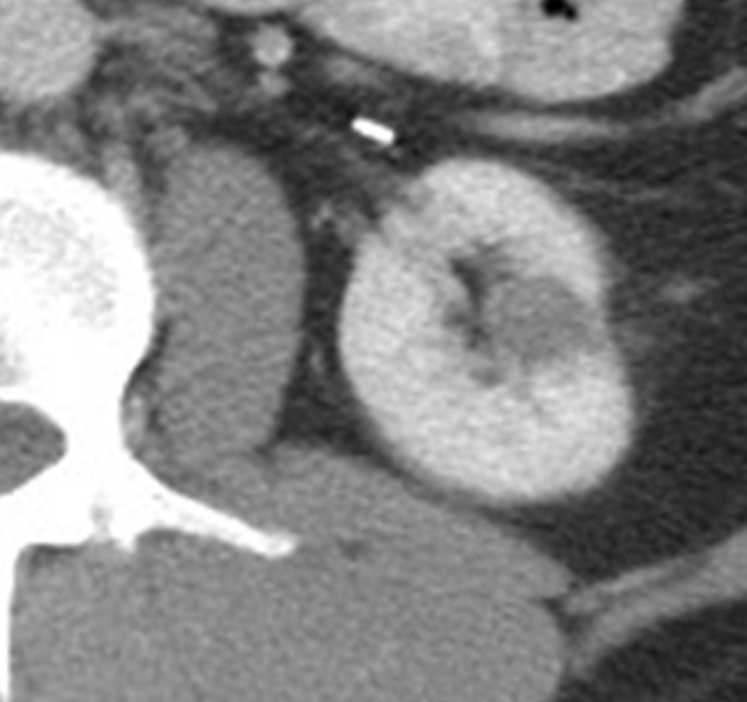
Expected evolution of imaging findings prior to and following percutaneous RF ablation. (a) Axial contrast-enhanced CT scan shows 1.5-cm enhancing RCC abutting left renal collecting system. (b) Axial intraprocedural nonenhanced CTscan shows electrode (arrow) within the tumor. (c) Axial contrast-enhanced CT scan 2 months after ablation shows trace left perinephric fat stranding and nonenhancement in the ablation zone. Nodularity within left perinephric fat (arrows), when observed along the electrode track, may mimic tumor seeding. (d) Axial contrast-enhanced CT scan 6 months after percutaneous RF ablation shows decreased size of ablation zone and decreased left perinephric fat stranding. Axial contrast-enhanced CT scans (e) 12 and (f) 18 months after ablation show continued decrease in size of ablation zone and continued decrease in left perinephric fat stranding and nodularity.