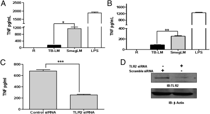
Fig. 1.
LM from M.tb stimulates minimal TNF production, whereas LM from M. smegmatis stimulates robust TNF production depending on TLR2. (A and B) MDM monolayers (A) or THP-1 cells (B) were incubated with TB-LM (5 μg/mL), SmegLM (5 μg/mL), or LPS as a positive control for 24 h. Cell-free culture supernatants were analyzed for TNF production by ELISA. The results in A and B are cumulative data from five and three experiments, respectively, each performed in triplicate (*P < 0.01; **P < 0.001). (C) MDMs were transfected with TLR2 siRNA or scramble siRNA (control) and plated in RPMI containing 20% autologous serum. After 24 h, cells were washed and stimulated with SmegLM (5 μg/mL) for 24 h. Cell culture supernatants were collected and analyzed for TNF production by ELISA (***P < 0.0001). (D) The cell lysates were examined for TLR2 and β−actin expression by Western blot analysis. The graph and Western blot are representative of three experiments.

