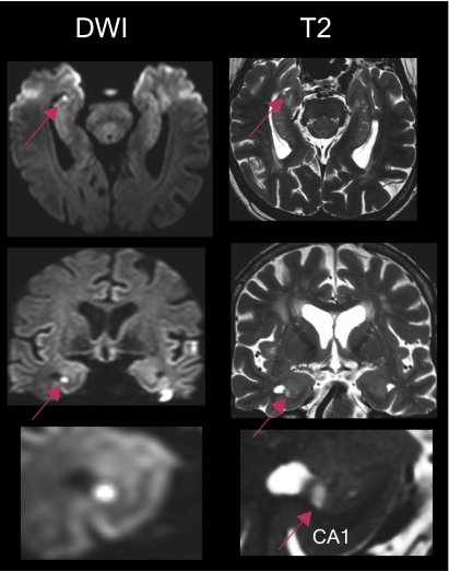
Fig. 5.
Whole-brain 3T MRI of one patient showing a focal lesion in the head of the right hippocampus. The lesion in the diffusion-weighted imaging correlates with the lesion in T2-weighted sequences (red arrow). The lesion as seen in the diffusion-weighted image can be clearly differentiated from the hyperintense susceptibility artifacts at the skull base. Imaging in the coronal plane show that the lesion is confined to the CA1 sector of the cornu ammonis (Bottom, red arrow).

