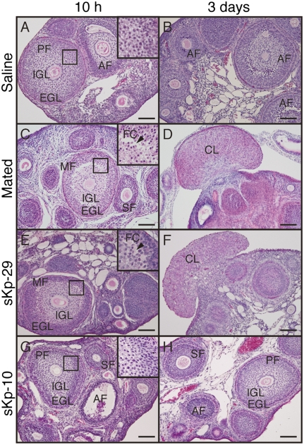
Fig. 4.
Effects of sKp-29 and sKp-10 on follicular development and corpus luteum formation. Photomicrographs of ovaries with H&E staining in animals that were mated (C and D) or injected with saline (A and B), sKp29 (E and F), or sKp-10 (G and H). Photographs were taken 10 h (A, C, E, and G) or 3 d (B, D, F, and H) after the onset of mating or injection. Mated and sKp-29–injected shrews showed slit-like follicular cavity (arrowheads) in ovarian follicles at 10 h and fungiform corpora lutea in the ovary at 3 d. Insets show the boxed area in each panel at higher magnification. AF, atretic follicle; CL, corpus luteum; EGL, extra granulosa layer; FC, follicular cavity; IGL, inner granulosa layer; MF, mature follicle; PF, premature follicle; SF, secondary follicle. (Scale bars: 100 μm.)

