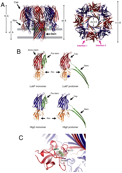
Fig. 1.
Overall octameric pore structure of γHL. (A) Side and top views of the heptamer. LukF and Hlg2 are shown in red and blue, respectively. MPD molecules bound with LukF are shown as cyan spheres. The aromatic side chains located around the putative membrane surface are shown as green sticks. The putative membrane region is also shown in gray. (B) Structures of the protomers of LukF (Upper) and Hlg2 (Lower). Monomeric structures of each molecule are also shown. Red, cap of LukF; blue, cap of Hlg2; green, stem (prestem in monomers); orange, rim; cyan, amino-latch. Blue spheres represent MPD bound with LukF protomer. (C) Close-up view of the MPD binding site. The bound MPD, Trp177, and Arg198 are shown as sticks. The Fo-Fc map (contoured at 1.5σ) around the MPD is also shown.

