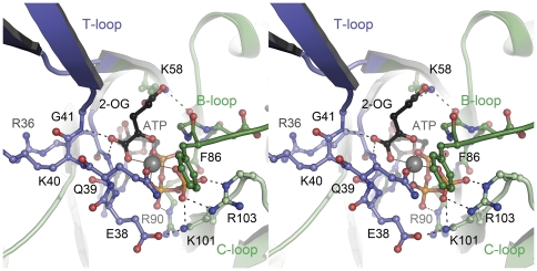
Figure 3. Binding mode of the ligands ATP, Mg2+ and 2-oxoglutarate to Af-GlnK3.
The stereo image shows a view into the ligand-binding cleft located at the interface of two monomers, one of which (dark green) provides the T-loop (blue) and B-loop regions to the binding site, the other monomer (light green) the C-loop. The Mg2+ ion (grey sphere) shows octahedral coordination by all three phosphate groups of ATP, by the á-carboxy and á-keto functions of 2-oxoglutarate and by the ã-amido oxygen atom of residue Q39.

