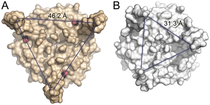
Figure 4. Structural consequences of ligand binding to GlnK proteins.
(A) With the ligand 2-OG (shown in CPK representation) placed in a wedge-like manner at the base, the T-loops of Af-GlnK3 are pried apart in a locked conformation. In the trimer, residues R47 of the monomers are 46.2 Å apart, a distance too large to be able to insert into the substrate channels of the cognate ammonium transporter. (B) Structure of the E. coli ortholog GlnK as seen in complex with the ammonium transporter AmtB (PDB-ID 2NS1) [27]. The T-loops are ordered and are positioned to fit the substrate channels of the transporter trimer, at a distance of 31.3 Å between residues R47.

