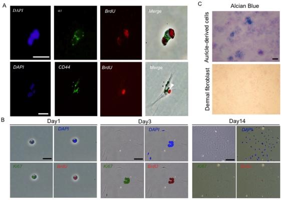
Figure 5. In vitro characterization of CD44+ integrin-α5+ LRCs.
(A) Immunocytochemistry of LRCs in vitro. Integrin-α5 and CD44 were co-expressed in BrdU-labeled cells. CD44 staining: scale bar = 20 µm; integrin-α5 staining: scale bar = 40 µm. Original magnification: ×200. (B) Colony formation of LRCs. Cells from 24-week-old mice, which were injected with BrdU as E17 to E19 fetuses, were harvested from mice auricle following collagenase digestion. Colony assay was performed to examine the clonogenicity of LRCs. Cells were stained at 1, 3 and 14 day after plated. Clonal colonies were stained with antibodies against DAPI, Ki-67 and BrdU. Scale bars = 20 µm or 100 µm (C) Alcian blue staining of cells isolated from auricular cartilage or dermal fibroblasts. Scale bar = 200 µm.

