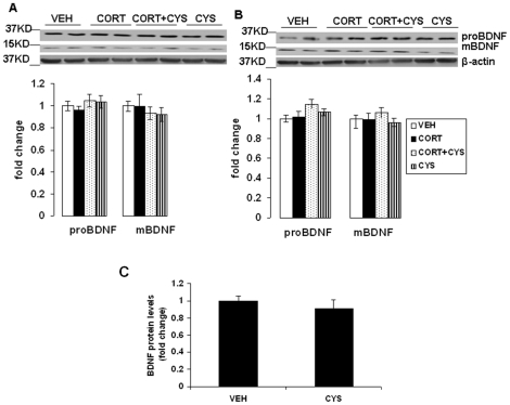
Figure 6. Effects of cysteamine in chronic corticosterone-treated mice on proBDNF and mature BDNF (mBDNF) protein levels in the frontal cortex and hippocampus.
CD-1 male mice were treated for 7 weeks with vehicle (0.45% hydroxypropyl-β-cyclodextrin) or corticosterone (CORT; 35 ug/ml) in the presence or absence of cysteamine (CYS; 150 mg/kg/day) during the last three weeks of corticosterone treatment. proBDNF and mBDNF protein levels were determined in the (A) frontal cortex and (B) hippocampus by Western blot analysis. The upper panels shows a representative autoradiogram of proBDNF and mBDNF and the lower panel represents the fold change in optical density values normalized to vehicle-treated controls. β-actin was used as a protein loading control. Values are mean ± SE (n = 6 mice per group). (C) BDNF protein levels as measured by ELISA in frontal cortex samples from mice treated with vehicle or cysteamine for 3 weeks as above. Data represent the fold change in BDNF protein levels (pg/mg protein) normalized to vehicle-treated controls. Values are mean ± SE (n = 5 mice per group).

