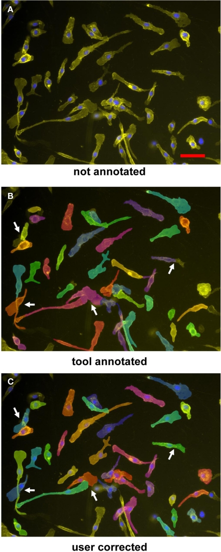
Figure 2.
Segmentation and annotation of macrophages with two-step segmentation software. Fluorescence microscopy images of CD11b–APC- and DAPI-stained macrophages were uploaded into the software (A), nuclear segmentation and contact area annotation was performed by the software tool (B), and finally checked and corrected by the user (C). Arrowheads indicate shrinkage effects; arrows point to overlapping cells necessitating manual editing of the annotation. Cells were imaged by a 20× objective. Scale bar represents 50 μm.

