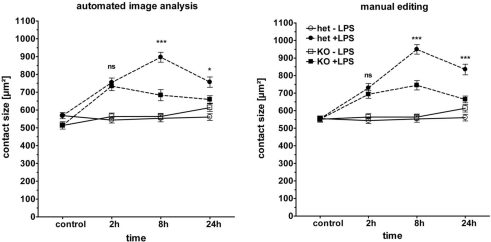
Figure 5.
Phenotype of Myd88−/− macrophages in cell spreading. BMM from Myd88± and Myd88−/− mice were plated and stimulated as described in Figure 3. Macrophage spreading was analyzed by automated image analysis (left panel) and manual editing (right panel). Shown are mean and SEM from one representative experiment of two performed. LPS (closed symbols), media control (open symbol), Myd88± (circles), Myd88−/− (squares). Statistical significance refers to Myd88± compared to Myd88−/− genotypes. ns = not significant; *** p < 0.0001.

