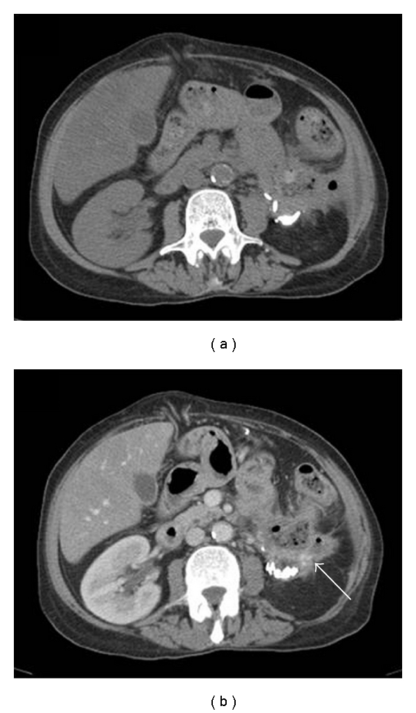
Figure 2.

CT scan of the abdomen and pelvis. (a) Imaging without i.v. contrast. Stranding and thickening of the soft tissue immediately below the lower pole of the left kidney and around the splenic flexure. (b) Imaging after i.v. contrast application. Stranding and thickening of the soft tissue around the kidney appears more prominent (arrow).
