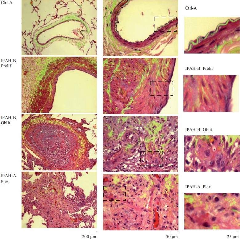
Figure 1.

Representative histopathologic changes observed in idiopathic pulmonary hypertension. Sections of human lungs (Ctrl-A, IPAH-A and IPAH-B) were stained using H&E and imaged using a ×40 objective in visible light. Elastin autofl uoresence was simultaneously imaged in green and the visible light and autofluoresence images merged. Representative images showing neointimal proliferation (Prolif), obliterative (Oblit) and plexiform (Plex) lesions are illustrated. Side sets on the right show higher magnifi cation views of the boxed areas within panels in the middle column. (Adapted from ref. 2.)
