
Figure 10.
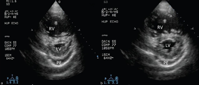
Short axis image at the midventricular level. Note the difference in the degree of septal flattening between systole (A) and diastole (B). The predominant systolic septal bowing is typical for PAH and other pulmonary vascular disease states. Also note the mild-moderate, circumferential pericardial effusion (PE), a common accompanying feature of the decompensated right ventricle.