
Figure 13.
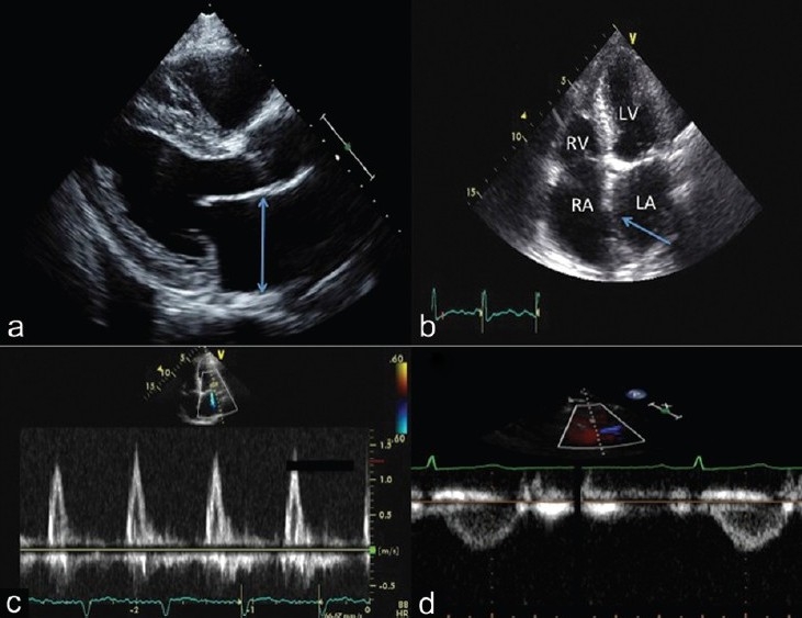
Integrated 2-D and Doppler echocardiographic assessment of a patient with PVH. Note the left atrial enlargement (panel A and B) and the left to right atrial septal bowing (panel B, arrow). Panel C demonstrates a restrictive transmitral flow pattern. The pulsed wave Doppler of the RVOT reveals no notching in the FVE (panel D), consistent with a normal pulmonary vascular resistance.