
Figure 14.
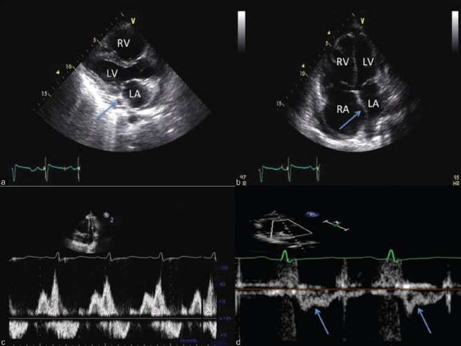
Integrated 2-D and Doppler echocardiographic assessment of a patient with PAH. Note normal left atrial size with a dilated coronary sinus (panel A, arrow) and right to left atrial septal bowing (panel B). Panel C demonstrates a Grade I diastolic dysfunction pattern (panel C) and the pulsed wave Doppler profile in the RVOT reveals mid-systolic notching (panel D, arrows), consistent with a very high pulmonary vascular resistance.