
Figure 7.
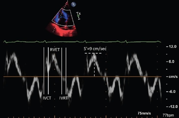
Right ventricular Tissue Doppler imaging along the basal long axis of the right ventricular free wall. The longitudinal shortening is labeled S’ and is measured at 9cm/sec. Right ventricular myocardial performance index can be obtained from the isovolumic contraction time (IVCT), relaxation time (IVRT), and ejection time (RVET).