
Figure 8.
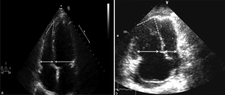
Apical four chamber view. Panel A demonstrates normal RV:LV size ratio (<1.0) and shape, with a preserved acute angle of the RV apex in a patient either without PH or with pulmonary venous hypertension. Panel B represents a patient with PAH or another form of PH with pulmonary vascular disease. Note the RV:LV ratio is increased (>1.0), the angle of the apex is less acute, and the RV is apex-sharing with the LV.