Fig. 3A,B.

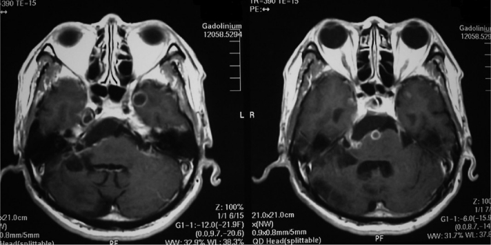
On a gadolinium magnetic resonance image, a tumor was visualized in the right cerebello-pontine angle, with a cyst wall in the bilateral temporal lobes.

On a gadolinium magnetic resonance image, a tumor was visualized in the right cerebello-pontine angle, with a cyst wall in the bilateral temporal lobes.