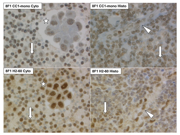
Figure 4.
Cyto-histologic comparison of anti-ERCC1 immunoreactivity using the protocol 8F1 CC1-mono (top) or 8F1 H2-60 (bottom). Left: Pleural effusion sediment of lung adenocarcinoma. Right: thoracic lymph node. Arrow: Lymphocyte. Arrowhead: Streak of endothelial cells. Asterisk: Tumor cell cluster. 400 × original magnification.

