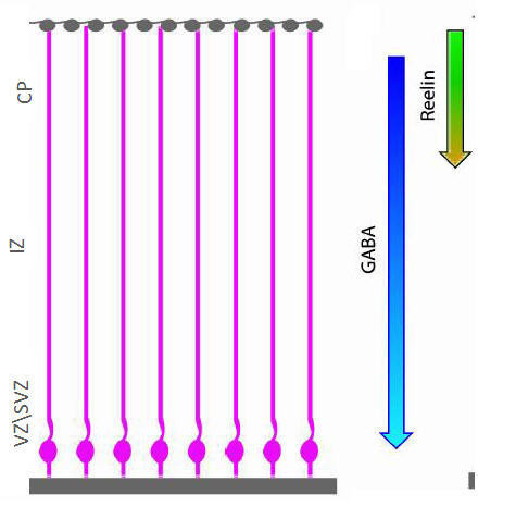
Figure 7.

Illustration of the simulated cortical cross-section. Glial cells, whose nucleus positions lie at the bottom of the cross-section and their fiber is stretched to the surface of the cortex (of the cross-section). Reelin and GABA factors are present at densities that lie in a gradient from top to bottom of the cross-section.
