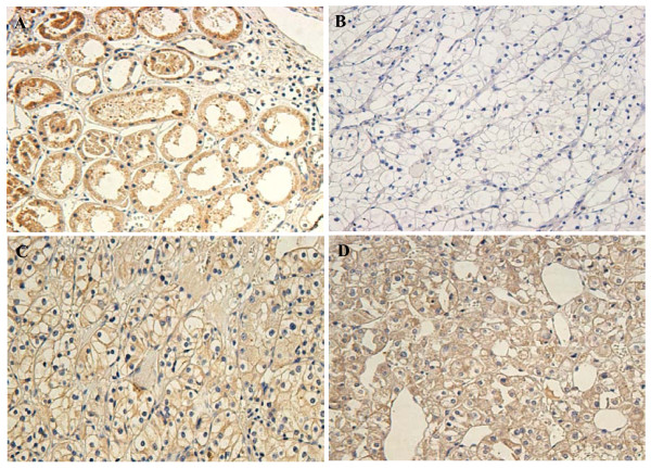
Figure 3.
Immunohistochemical analysis of the expression of DUSP-9 protein. DUSP-9 is mainly localized within the nuclei and cytoplasmic. Immunostaining of the adjacent normal tissue samples(A) and the ccRCC tumor tissue samples(B) showed a sharp contrast between the negatively stained infiltrative tumorous area.(B): Negative or weak DUSP-9 staining in cancerous tissue (400×). (C): Moderate DUSP-9 staining in cancerous tissue(400×). (D): Strong DUSP-9 staining in most of tumor cells (400×).

