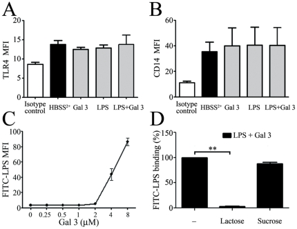
Figure 4. Gal 3 enhances LPS binding to the neutrophil surface.
A and B: PMNs (2×10⁶/mL) were stimulated for 45 min with Gal 3 (0.4 µM) and E. coli LPS (1 µg/ml), preincubated as in figure 3. TLR4 (A) and CD14 (B) expressions were determined by flow cytometry. The results are expressed as mean fluorescence intensity (MFI) (n = 3). C: FITC-labeled E. coli LPS (12.5 µg/mL) was preincubated with increasing molar concentrations of Gal 3 (from 0.25 to 8 µM) and then added to PMNs (10×106/mL) for additional 20 min incubation at 37°C with (n = 3) D. Gal 3 was treated with 12.5 mM lactose or sucrose for 15 min before being incubated with FITC-LPS (12.5 µg/mL) and added to PMNs (10×106/mL). The binding of FITC-LPS was determined by flow cytometry and expressed as mean fluorescence intensity (MFI).

