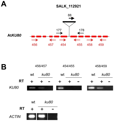
Figure 4. T-DNA insertion and expression in ku80 mutant.
(A) Position of the T-DNA insertion in AtKU80. The structure of the AtKU80 gene is represented by shaded boxes (exons) and thin lines (introns). The T-DNA insertion position is indicated. Each primer pair used to characterize the mutant by PCR are indicated in black and primer pairs used for RT-PCR analyses are given in red; their localization is correct but not to scale. (B) RT-PCR analysis of AtKU80 transcripts in ku80-/- mutant plants. RNA, extracted from floral buds of wild-type or ku mutant plants was reverse-transcribed. Double-stranded cDNAs were amplified by RT-PCR, performed with three different primer pairs: 5′ or 3′ to the T-DNA and flanking the T-DNA insertion. For primer positions, see above (Figure 4A). The constitutive ACTIN gene was used as a control.

