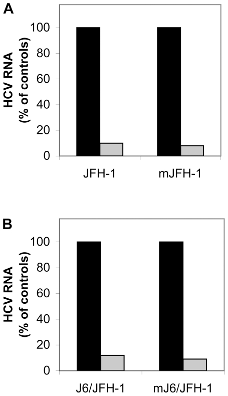
Figure 1. LPL inhibits cell infection by the JFH-1 and J6/JFH-1 strains produced in vitro and in vivo in a chimeric uPA-SCID mouse model.
The HCVcc strains JFH-1 (A) and J6/JFH-1 (B) were produced in the Huh7.5 hepatoma cell line. Cells were incubated with (or without) LPL for 30 min at 4°C and then with virus preparations for 2 h at 37°C to allow infection. RNA was extracted from cells 24 h post infection and HCV RNA was quantified by RT-qPCR. The data obtained were normalized with respect to levels of GADPH. The mJFH-1 (A) and mJ6/JFH-1 (B) correspond to HCVcc strains produced in chimeric uPA-SCID mice into which we transplanted human hepatocytes. Serum samples collected from infected mice were pooled and their capacity to infect Huh7.5 cells was assessed in the presence and absence of LPL, as outlined above. Cells infected in the absence (black bar) and in the presence of LPL (gray bar). The data are expressed as the amount of HCV RNA detected in cells infected in the presence of LPL as compared with the amount of HCV RNA in cells infected in the absence of LPL, expressed as a percentage.

