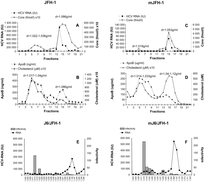
Figure 2. Iodixanol gradient analysis of the JFH-1 and J6/JFH-1 strains produced in vitro and in vivo.
The supernatants from infected Huh7.5 cells producing JFH-1 (JFH-1, shown in A and B) and J6/JFH-1 (shown in E) were subjected to isopycnic centrifugation through iodixanol gradients, as described in Materials and Methods. Pooled serum samples from the chimeric uPA-SCID mice were also subjected to centrifugation on the same type of gradient. Representative profiles are shown in C and D for mice inoculated with JFH-1 (mJFH-1) and in F for mice inoculated with J6/JFH-1 (mJ6/JFH-1). HCV core antigen in gradient fractions was quantified by ELISA, HCV RNA was quantified by RT-qPCR, and ApoB and cholesterol were determined by ELISA. Infectivity for fractionated J6/JFH-1 (representative for both strains) grown in Huh7.5 cells is shown in E and that for the corresponding mouse serum (mJ6/JFH-1) is shown in F. The fractions (25 µl) were used to infect Huh7.5 cells. Cells were incubated for 48 h at 37°C; total RNA was then extracted and HCV-RNA levels were quantified by RT-qPCR. The results were normalized, taking into account the initial HCV-RNA content in each sample analyzed, as determined by RT-qPCR, and are expressed as a ratio of these two values.

