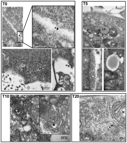
Figure 7. Immunoelectron microscopy of the infection of Huh 7.5 cells with HCV in the absence of LPL.
The viral preparation (JFH-1) was concentrated by centrifugation through a sucrose cushion and incubated with Huh7.5 cells at 4°C (T0), before transfer to 37°C and incubation for a further 5 (T5), 10 (T10), 15 (T15) or 20 (T20) min. Cells collected at all these time points were washed, fixed and stained with monoclonal antibodies, followed by secondary, colloidal gold-labeled anti-mouse IgG (see Materials and Methods section for details). T0 and T5, immunogold labeling of HCV E2 envelope glycoprotein with monoclonal AP-33 antibody; T10 and T20, immunogold labeling of HCV core protein with monoclonal ACAP-27 antibody. Asterisks indicate the presence of one silver-enhanced gold particle. ER, endoplasmic reticulum; LD, lipid droplet; M, mitochondrion; Nu, nucleus.

