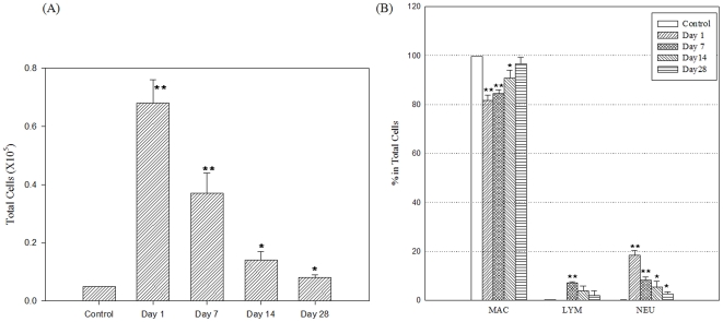
Figure 3. Changes in the cell distribution in BAL fluid after instillation of DEPs (n = 4).
Mice were administered a single intratracheal instillation of DEPs at a dose of 10 mg/kg and then killed on the designated day (1, 7, 14, or 28 days after exposure). The cells in the BAL fluid were quantified by hemocytometric counting (A), and the distributions of alveolar macrophages, neutrophils, and lymphocytes were assessed on the basis of their characteristic cell shapes (B). The cell number in each group was expressed as mean ± SD. *; P<0.05, **; P<0.01.

