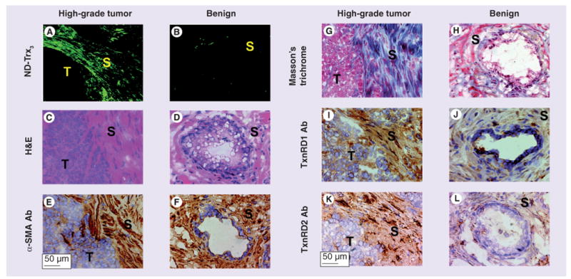
Figure 6. Binding of thioredoxin-targeted ND-Trx3 mirrors binding of antibodies to thioredoxin reductases 1 and 2.

Sections (5-μm thick) of freshly frozen tissue specimens obtained after resection from patients with biopsy-proven prostate cancer (A) or benign prostatic hyperplasia (BPH) (B) were stained with ND-Trx3 or immunostained with antibodies to α-SMA, TxnRD1 or TxnRD2. Serial sections of each case were stained with H&E to confirm the presence of cancer (C), BPH (D) and their cellular characteristics in the particular section. Anti-α-SMA antibody showed strong reactivity with stromal cells in cancer (reddish brown staining [E]) and BPH tissue specimens (reddish brown staining [F]). Masson's trichrome staining was strongly positive for reactive stromal cells near the tumor lesion (G) but not with the stromal cells in BPH specimens (H). Trichrome-stained stromal cells (G) also showed reactivity with anti-TxnRD1 antibody (reddish brown staining [I]) and ND-Trx3 (fluorescence [A]) in cancer, but not BPH tissue specimens (absence of reddish brown staining [J] and fluorescence [B]). A similar specificity and pattern of α-SMA-positive stromal cells with anti-TxnRD2 antibody (reddish brown staining [K]) and ND-Trx3 (fluorescence [A]) was observed in cancer or BPH tissue specimens [L & B]). The sections were counterstained with Harris's hematoxylin (blue nuclear staining). Original magnification: 100×. Tumor (T) and stromal (S) regions are indicated in each panel.
Ab: Antibody; α-SMA: α-smooth muscle actin; H&E: Hematoxylin and eosin; ND-Trx3: Nanodevice; S: Stromal region; T: Tissue region; TxnRD: Thioredoxin reductase.
