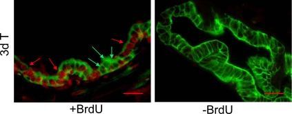
Fig. 4.
Proliferation of GFP-labeled luminal epithelial cells after androgen replacement. BrdU was injected 3 d and 4 d after testosterone replacement, and the prostate was isolated 4 h after the second injection (two-cycle group as described in Fig. 3). Red arrows indicate BrdU-labeled, GFP-positive cells (top panel). Green arrows indicate BrdU-negative, GFP-positive cells. Animals without BrdU injection were processed in parallel as negative control. Scale bars, 25 μm. T, Testosterone.

