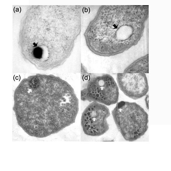
Figure 1.
Electron micrographs thin sections of Agrobacterium tumefaciens(a &b) and Methanosarcina acetivorans (c & d). In panel (a), the arrow shows the partially filled acidocalcisome of A. tumefaciens containing electron dense material. In panel (b), the arrow shows an empty A. tumefaciens acidocalcisome. In panel (c), the arrow shows the electron dense volutin granule of M. acetivorans. In panel (d), the arrows show empty, partially, and completely filled volutin granules of M. acetivorans.

