
Fig. 4.
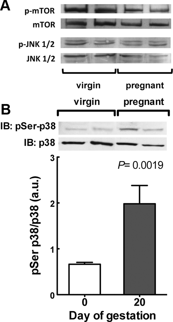
p38 MAPK activation is increased in adipose tissue at late gestation. A, Phosphorylation of mTOR and JNK1/2. Representative immunoblots for phosphorylated and total proteins of two independent experiments. B, Phosphorylation of p38 MAPK. The graph shows the levels of phosphorylated p38 MAPK, normalized to the total amount of p38 MAPK protein, of six independent experiments. A representative immunoblot (IB) for phosphorylated and total protein is shown in the graph.