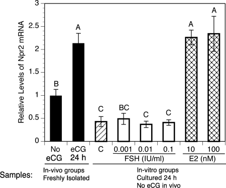
Fig. 3.
Effect of FSH and estradiol in vitro on levels of Npr2 mRNA in cumulus cells of COCs isolated from mice that were not stimulated with eCG. The mean value in COCs freshly isolated from mice not stimulated by eCG was set at 1, and other levels are shown relative to it. Bars show the mean ± sem of three independent samples. The solid bars compare levels in COCs freshly isolated from unstimulated mice to those taken from mice 24 h after eCG. The single hatched bar (C) shows the relative level in control COCs cultured 24 h without either FSH or estradiol (E2). White bars show relative levels in COCs cultured in various concentrations of FSH for 24 h. Cross hatched bars show relative levels in COCs cultured 24 h in 10 or 100 nm estradiol. Relative expression levels not connected by the same letter are significantly different (P < 0.05).

