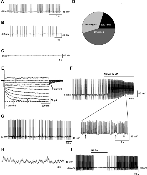
Fig. 4.
Electrophysiological characteristics of arcuate Kiss1 neurons in oil-treated OVX Kiss1-CreGFP mice using whole-cell patch recording. Kiss1 neurons in the ARC (of the female) rest at −63.8 ± 2.3 mV (n = 20). A–C, Representative traces of action potentials recorded from arcuate Kiss1 neurons showing tonic (A), irregular (B), and silent (C) firing patterns. D, Summary pie chart of the firing pattern distribution in ARC Kiss1 neurons (n = 20). E–H, Endogenous conductances and NMDA-induced burst firing of an ARC Kiss1 neuron. E, Ensemble of currents in response to depolarizing/hyperpolarizing steps from −50 to −140 mV illustrating the expression of a hyperpolarization-activated cation current (h-current) and a T-type Ca2+ current (arrow) in a representative Kiss1 neuron. Vhold = −60 mV. F, Current clamp recording in an ARC Kiss1 neuron showing the response to NMDA (40 μm). The spiking in F was expanded to illustrate the pronounced effects of NMDA on burst firing activity of Kiss1 neurons with an ensemble of spikes riding on top of low-threshold spikes (arrows). G, In a current clamped state close to the resting membrane potential, one can see the bursting activity in the presence of NMDA. H, In the presence of tetrodotoxin to block the fast Na+ spikes, one can clearly see the membrane oscillations (up and down states) induced by NMDA. Eighty percent of ARC Kiss1 neurons expressed the endogenous conductances (h-current, T-current) that are critical for generating burst firing activity. Although ARC Kiss1 neurons did not exhibit spontaneous burst firing activity, glutamate (NMDA) was able to induce burst firing activity in all of the cells. I, GABA (100 μm) inhibited firing in another arcuate kisspeptin neuron. Drugs were rapidly perfused into the bath as a 100-μl bolus.

