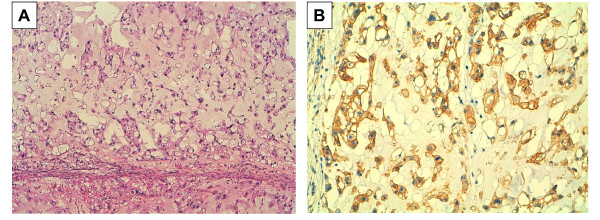
Figure 2.
Chordoma and EGFR overexpression. A/H&E staining showing round cells with vacuolated cytoplasm arranged in cord-like fashion in a myxoid stroma (original magnification × 100). B/Immunohistochemistry with anti-EGFR antibody showing strong membranous and cytoplasmic staining of tumor cells (original magnification × 200; positivity appears in brown).

