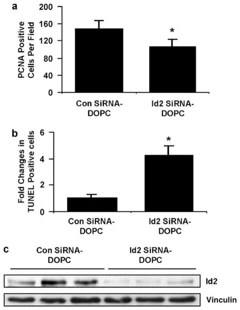
Figure 6.
Effect of in vivo administration of liposomal-conjugated siRNA to Id2 on CRC tumor growth in the liver. (a) Graphical plot of immunohistochemistry (IHC) staining for proliferating cell nuclear antigen (PCNA) in tumor sections from samples in Figure 5c. Treatment groups; control liposome (control siRNA–DOPC) and Id2-targeted liposome (Id2 siRNA–DOPC). *P = 0.02 between treatment groups. (b) Graphical representation of ‘fold’ change in TUNEL-positive cells (apoptotic cells) in tumor sections from control-siRNA- and Id2-siRNA-treated mice observed in five fields at ×20. Average *P = 0.002 between treatment groups. (c) Western analysis of tumor homogenates for Id2 expression in liposomal treatment groups from Figure 5c; control liposome (control siRNA–DOPC) and Id2-targeted liposome (Id2 siRNA–DOPC). Id2, inhibitor of DNA-bind-2; siRNA, small interfering RNA; DOPC, 1,2-dioleoyl-sn-glycero-3-phosphatidylcholine; TUNEL, TdT-mediated UTP nick-end labeling.

