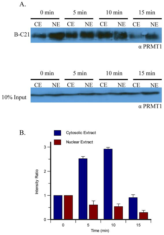
Figure 6.
Labeling MCF-7 Cytoplasmic and Nuclear Extracts Following Estrogen Stimulation. A. MCF-7 cells were stimulated with estrogen for the indicated amount of time and then the cytoplasmic and nuclear extracts were incubated with B-C21 and bound to streptavidin-agarose beads overnight. Bound proteins were eluted from the beads, separated by SDS-PAGE and visualized by western blot analysis using an anti-PRMT1 antibody (upper panel). To demonstrate equal protein loading, a 10% loading control was loaded onto a separate gel and visualized by western blot analysis using the same antibody (lower panel). Representative data from one of three experiments is depicted. B. Quantification of the western blots in panel A.

