Figure 1.
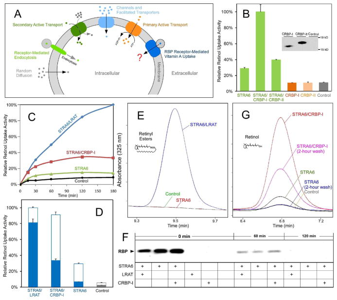
STRA6 couples cellular vitamin A uptake to intracellular storage. (A) Schematic diagram comparing known cellular uptake mechanisms (e.g., receptor-mediated endocytosis, primary and secondary active transport, channels, facilitated transport, and random diffusion) and RBP receptor-mediated cellular vitamin A uptake. (B) CRBP-I is much more effective than CRBP-II in coupling with STRA6 to take up 3H-retinol from 3H-retinol/RBP. Both CRBP-I and CRBP-II were tagged with 6X His tag at the C-terminus and their expression in transfected cells was detected with anti-His tag antibody to compare their expression levels. (C) Comparison of the time-dependent accumulation of 3H-retinol for STRA6/LRAT, STRA6/CRBP-I, STRA6 only cells, and control untransfected cells after uptake from 3H-retinol/RBP. The highest 3H-retinol uptake activity of STRA6/LRAT cells is defined as 100%. (D) Removal of cell surface bound 3H-retinol/RBP by excessive unlabeled holo-RBP after 3H-retinol uptake from 3H-retinol-RBP. Unlike STRA6/LRAT cells, most 3H-retinol associated with STRA6 cells (signal above the background of untransfected cells) represents 3H-retinol/RBP bound to the cell surface. Unfilled columns represent cell surface-bound 3H-retinol in the form of 3H-retinol/RBP. Filled columns represent cellular 3H-retinol uptake. (E) HPLC analysis of retinyl ester formation by STRA6/LRAT cells, STRA6-only cells, and control untransfected cells using human serum as the source of holo-RBP. Peaks representing retinyl esters are shown. Given the role of LRAT in retinyl ester formation, STRA6-only and control cells have no detectable retinyl esters. (F) Analysis of the dissociation of RBP bound to cells transfected with STRA6, STRA6/LRAT, STRA6/CRBP-I and control cells. After cells were incubated with holo-RBP for one hour, unbound RBP was removed by washing cells with PBS. After further incubation in serum free media (SFM) for 0, 60, or 120 minutes, RBP associated with cells was analyzed by Western blot. RBP completely dissociated from STRA6 after two hours of incubation in SFM. (G) HPLC analysis of retinol uptake from human serum by cells transfected by STRA6/CRBP-I, STRA6, and control cells. Two hours of incubation in SFM removed cell surface bound RBP. Peaks representing retinol are shown. One unit on the Y axis represents 1 mAU for E and G.

