Figure 6.
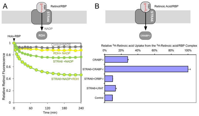
LRAT and CRBP-I-independent enhancement of STRA6’s substrate uptake. Schematic diagram of each experiment is shown on the top. (A) Retinol fluorescence assay (4 μM holo-RBP added at 0 min) showing that RDH significantly enhanced STRA6-catalyzed retinol release. NADP (0.2 mM) was added to all reactions. (B) CRABP-I, but not CRBP-I or LRAT, couples with STRA6 for cellular 3H-retinoic acid uptake from 3H-retinoic acid/RBP. The activity of STRA6/CRABP-I transfected cell is defined as 100%. Activity in CRABP-I-only cells was due to non-specific release of retinoic acid from RBP into cells. The inability of CRBP-I and LRAT to couple to STRA6 in retinoic acid uptake is consistent with the facts that CRBP-I does not bind retinoic acid and retinoic acid is not a substrate for LRAT. A schematic diagram for each experiment is drawn on the top of each experiment.

